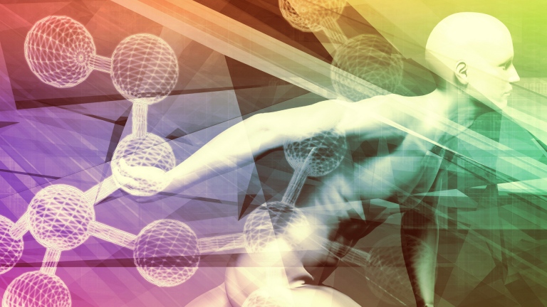
Regrowth Neurons are also called Neuro-Regeneration.
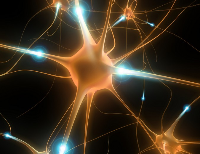
Regrowth neurons… also called Neuro-Regeneration, is one of the most complicated issues in medicine/science. The subject matter concerns the repair and regrowth of nerve cells called neurons. The long body of the neuron is called the Axon. This issue is divided into two imperative groups….
The brain and spinal cord characterize the peripheral Nervous System (PNS) and the Central Nervous System (CNS). PNS has a method of regeneration to the injured neurons followed by the quick release of white blood cells to fight infection. Schwann cells release neurotrophic factors, which enhance the regrowth of PNS Regrowth Neurons in several ways. The Schwann cells prevent too much cytotoxicity, which is generally part of the healing process but elicits too much inflammation for regeneration to proceed normally.
Schwann cells also set up tubes alongside the damaged neuron. These tubes are even connected to the injured degraded PNS neuron, and the pipe releases a host of healing biochemicals to stop neural degradation and provide healing repair to the affected injured neuron. A host of outside interventions can make this process go faster. However, I will save them for Regeneration of the brain and spinal cord, which, please remember, is still thought in many universities, even taught to this day, that CNS Regrowth Neurons cannot regenerate!
Regeneration and Repair of the Central Nervous System (Brain and Spinal Cord. Two types of cells are meant to serve and protect CNS Regrowth Neurons from injury. They are called Astrocytes and Glial cells. It may be hard to believe, but there are much more astrocytes in the brain than there are brain Neurons, and some 15 to 18 billion brain Regrowth Neurons. Normally, after an injury to brain neurons, astrocytes support Regrowth Neurons by providing antioxidant protection.
The Good, the Bad, and the Ugly
While the Astrocytes bring in many antioxidants to prevent free radical oxidative toxic damage to the brain Regrowth Neurons, they also over-react and allow for too much inflammation and cytotoxins from white blood cell production of Interleukin-2 and 6. This is called the up-regulation of immune cell activity. It can destroy brain neurons faster than the initial injury to a brain or spinal neurons.
This process brings about even more inflammation to try and carry away all dead cells, including neurons and astrocytes, as well as white blood cell waste deposits. Unfortunately, the brain is limited in size, and too much inflammation causes intracranial pressure to the point where brain infarction is possible without making a surgical opening in the skull to release the stress….that is the bad and the ugly.
How do we solve the above dire situation and bring about brain cell neuron regeneration?
In some cases, like bacterial or viral Encephalitis of the brain and spinal cord, many Regrowth Neurons can be killed or injured due to massive white blood cell proliferation. The white blood cells destroy the bacteria and virus that causes Encephalitis. Still, the situation is so dramatic that for the brain to survive the infection-killing neurons, the message which signals the white blood cells to enter the brain becomes so up-regulated that it cannot down-regulate to a normal response, thus further killing both infected Regrowth Neurons and the infected brain cells both. Many people die from this berserk super up-regulated immune response stuck on full throttle. It is a vicious, repetitive cycle.
We can stop this death cycle by giving the patient large intravenous doses of Gamma Globulin. Gamma Globulin consists of every type of antibody the human body can make. Scientists are not exactly certain how and why this has such a profound normalizing effect immunologically, but it does. It is called Immuno-modulation to the normal set point. In both Stroke patients and infection-based Encephalitis of the brain, Immuno-Modulation to a normal set point is a must for neuron cell death to stop and for regeneration to begin.
Certain biochemical factors play a vital role in decreasing astrocytes! Cyclin Kinase decreases astrocyte proliferation, increasing neuron function and recovery. Caffeic acid, alpha-melanocyte-stimulating hormone, and cilostazol are all highly beneficial. They are considered essential to an improved condition by reducing astrocyte production. This treatment shows a decrease in neuron injury. The decline in astrocyte high production levels is associated with a more positive and improved outcome moving toward CNS neuron regeneration.
1984 I experienced a life/death illness called Herpetic Viral Encephalitis. Manhattan’s most elite doctors misdiagnosed it…virtually always fatal, I lived, and because the death rate is so nearly complete, I spent three years in bed and several more in a wheelchair. There was no internet, no smartphones, just a nearly destroyed brain with pain so blinding it felt like two ice picks were being run through both eyes… and trying to escape by pushing through the back of my skull. Luckily for me, the feelers I sent out daily… resulted in a call from Nobel Prize-winning doctor and scientist Dr. Rita Levi Montalcini, who won the Nobel Prize for discovering Embryonic Fetal Nerve Growth Factor in 1986. I was the first human to get shots of the Fetal Nerve Growth Factor in 1988 to grow back and regenerate my damaged brain Regrowth Neurons. Then this magnificent woman became my mentor. I still live speaking to her shadow every day as she passed over at the age of almost 104.
Rita Levi-Montalcini’s discovery of a protein called ‘fetal nerve growth factor,’‘
which fosters the growth of nerve fibers and plays a role in the brain and the immune system; it is one of the most important steps taken so far toward understanding how the very complex system of nerves is laid down and linked to the tissues in a developing embryo. Her account of the adventures leading to this discovery, for which she won a Nobel Prize in Physiology and Medicine in 1986, has a special contemporary interest.
We now know her discovery is how the brain grows all of its Regrowth Neurons when one is an embryo inside one’s mother’s womb until birth and sustains, protects, and regenerates brain Regrowth Neurons if one is fortunate to be born with a large amount of FNGF. Thank goodness she could scale it up through a deal with a large cutting-edge biotechnology lab in Montreal. They are the ONLY Lab in the world that makes Dr. Rita’s original formulations.
We at AAI are incredibly fortunate to have access to her formulations at all times.
We combined her fetal nerve growth factors with very youthful blood levels of HGH (human growth hormone) that would be normal for the average healthy 16-year-old. Eighteen months later, I was completely better in every way, brain regeneration-wise and in every other health manner. The addition of HGH was my inspiration as it has been known for many decades that it is HGH that mobilizes all of the fetal tissue growth factors, such as cardiac tissue growth factor…to do protein synthesis and thus the regeneration of the heart AND all other tissues contained within the human body.
Without youthful blood levels of HGH (say blood levels normal for a healthy 26-year-old to 30-year-old) and credible levels of fetal nerve growth factor, regrowth of Neurons is unlikely in the extreme, BUT with these safe and effective blood levels of both regrowth of every type of Neuron is probably in, and that brings amazing hope.
The only Regrowth we cannot master yet is CNS spinal cord cut in half…but the army medical scientists are getting very close. Imagine how old we would all look if we did not have adequate cell levels of epithelial (skin tissue growth factor) to regenerate our SKIN?
So, just like hormones, as we age, we lose our tissue growth factors…and if they cannot be replaced to the optimal level from exogenous sources, we will all be subject to the ravages of age-related illness. Gratefully, here at AAI, we have all critical tissue growth factors available to you, our beloved family of clients.
REFERENCES
- Lloyd-Jones D, Adams RJ, Brown TM, Carnethon M, Dai S, De Simone G, Ferguson TB, Ford E, Furie K, Gillespie C, Go A, Greenlund K, Haase N, Hailpern S, Ho PM, Howard V, Kissela B, Kittner S, Lackland D, Lisabeth L, Marelli A, McDermott MM, Meigs J, Mozaffarian D, Mussolino M, Nichol G, Roger VL, Rosamond W, Sacco R, Sorlie P, Roger VL, Thom T, Wasserthiel-Smoller S, Wong ND, Wylie-Rosett J. Heart disease and stroke statistics–2010 update: a report from the American Heart Association.Circulation. 2010;121:e46–e215. [PubMed]
- Blakeley JO, Llinas RH. Thrombolytic therapy for acute ischemic stroke. J. Neurol. Sci. 2007;261:55–62.[PubMed]
- Marder VJ, Jahan R, Gruber T, Goyal A, Arora V. Thrombolysis with plasmin: implications for stroke treatment. Stroke. 2010;41:S45–S49. [PMC free article] [PubMed]
- Bernard SA, Gray TW, Buist MD, Jones BM, Silvester W, Gutteridge G, Smith K. Treatment of comatose survivors of out-of-hospital cardiac arrest with induced hypothermia. N. Engl. J. Med.2002;346:557–563. [PubMed]
- Howells DW, Porritt MJ, Rewell SS, O’Collins V, Sena ES, van der Worp HB, Traystman RJ, Macleod MR. Different strokes for different folks: the rich diversity of animal models of focal cerebral ischemia. J. Cereb. Blood Flow Metab. 30:1412–1431. [PMC free article] [PubMed]
- Kettenmann H, Ramson BR. Neuroglia. New York: Oxford University Press; 1995.
- Nedergaard M, Ransom B, Goldman SA. New roles for astrocytes: redefining the functional architecture of the brain. Trends Neurosci. 2003;26:523–530. [PubMed]
- Sofroniew MV, Vinters HV. Astrocytes: biology and pathology. Acta Neuropathol. 2010;119:7–35.[PMC free article] [PubMed]
- Goldberg MP, Choi DW. Combined oxygen and glucose deprivation in cortical cell culture: calcium-dependent and calcium-independent mechanisms of neuronal injury. J. Neurosci. 1993;13:3510–3524.[PubMed]
- Swanson RA, Ying W, Kauppinen TM. Astrocyte influences on ischemic neuronal death. Curr. Mol. Med. 2004;4:193–205. [PubMed]
- Dringen R, Hirrlinger J. Glutathione pathways in the brain. Biol. Chem. 2003;384:505–516. [PubMed]
- Wilson JX. Antioxidant defense of the brain: a role for astrocytes. Can. J. Physiol. Pharmacol.1997;75:1149–1163. [PubMed]
- Rossi DJ, Brady JD, Mohr C. Astrocyte metabolism and signaling during brain ischemia. Nat. Neurosci.2007;10:1377–1386. [PubMed]
- Nedergaard M, Dirnagl U. Role of glial cells in cerebral ischemia. Glia. 2005;50:281–286. [PubMed]
- Ouyang YB, Voloboueva LA, Xu LJ, Giffard RG. Selective dysfunction of hippocampal CA1 astrocytes contributes to delayed neuronal damage after transient forebrain ischemia. J. Neurosci.2007;27:4253–4260. [PMC free article] [PubMed]
- Xu L, Emery JF, Ouyang YB, Voloboueva LA, Giffard RG. Astrocyte-targeted overexpression of Hsp72 or SOD2 reduces neuronal vulnerability to forebrain ischemia. Glia. 2010;58:1042–1049.[PMC free article] [PubMed]
- Anderson MF, Blomstrand F, Blomstrand C, Eriksson PS, Nilsson M. Astrocytes and stroke: networking for survival? Neurochem. Res. 2003;28:293–305. [PubMed]
- Giffard RG, Swanson RA. Ischemia-induced programmed cell death in astrocytes. Glia. 2005;50:299–306. [PubMed]
- Almeida A, Delgado-Esteban M, Bolanos JP, Medina JM. Oxygen and glucose deprivation induces mitochondrial dysfunction and oxidative stress in neurons but not in astrocytes in primary culture. J. Neurochem. 2002;81:207–217. [PubMed]
- Xu L, Sapolsky RM, Giffard RG. Differential sensitivity of murine astrocytes and Regrowth Neurons from different brain regions to injury. Exp. Neurol. 2001;169:416–424. [PubMed]
- Zhao G, Flavin MP. Differential sensitivity of rat hippocampal and cortical astrocytes to oxygen-glucose deprivation injury. Neurosci. Lett. 2000;285:177–180. [PubMed]
- Lukaszevicz AC, Sampaio N, Guegan C, Benchoua A, Couriaud C, Chevalier E, Sola B, Lacombe P, Onteniente B. High sensitivity of protoplasmic cortical astroglia to focal ischemia. J. Cereb. Blood Flow Metab. 2002;22:289–298. [PubMed]
- Bondarenko A, Chesler M. Rapid astrocyte death induced by transient hypoxia, acidosis, and extracellular ion shifts. Glia. 2001;34:134–142. [PubMed]
- Giffard RG, Monyer H, Choi DW. Selective vulnerability of cultured cortical glia to injury by extracellular acidosis. Brain Res. 1990;530:138–141. [PubMed]
- Swanson RA, Farrell K, Stein BA. Astrocyte energetics, function, and death under incomplete ischemia conditions: a glial death mechanism in the penumbra. Glia. 1997;21:142–153. [PubMed]
- Tombaugh GC, Sapolsky RM. Mechanistic distinctions between excitotoxic and acidotic hippocampal damage in an in vitromodel of ischemia. J. Cereb. Blood Flow Metab. 1990;10:527–535. [PubMed]
- Chen Y, Swanson RA. Astrocytes and brain injury. J. Cereb. Blood Flow Metab. 2003;23:137–149.[PubMed]
- Lee MH, Kim H, Kim SS, Lee TH, Lim BV, Chang HK, Jang MH, Shin MC, Shin MS, Kim CJ. Treadmill exercise suppresses ischemia-induced increment in apoptosis and cell proliferation in the hippocampal dentate gyrus of gerbils. Life Sci. 2003;73:2455–2465. [PubMed]
- Li Y, Chopp M, Zhang ZG, Zhang RL. Expression of the glial fibrillary acidic protein in areas of focal cerebral ischemia accompanies the neuronal expression of 72-kDa heat shock protein. J. Neurol. Sci.1995;128:134–142. [PubMed]
- Schroeter M, Schiene K, Kraemer M, Hagemann G, Weigel H, Eysel UT, Witte OW, Stoll G. Astroglial responses in photochemically induced focal ischemia of the rat cortex. Exp Brain Res. 1995;106:1–6.[PubMed]
- Van Beek J, Chan P, Bernaudin M, Petit E, MacKenzie ET, Fontaine M. Glial responses, clusterin, and complement in permanent focal cerebral ischemia in the mouse. Glia. 2000;31:39–50. [PubMed]
- Yamashita K, Vogel P, Fritze K, Back T, Hossmann KA, Wiessner C. Monitoring the temporal and spatial activation pattern of astrocytes in focal cerebral ischemia using in situ hybridization to GFAP mRNA: comparison with sgp-2 and hsp70 mRNA and the effect of glutamate receptor antagonists. Brain Res.1996;735:285–297. [PubMed]
- Liu D, Smith CL, Barone FC, Ellison JA, Lysko PG, Li K, Simpson IA. Astrocytic demise precedes delayed neuronal death in focal ischemic rat brains. Brain Res. Mol. Brain Res. 1999;68:29–41. [PubMed]
- Dugan LL, Bruno VM, Amagasu SM, Giffard RG. Glia modulates the response of murine cortical Regrowth Neurons to excitotoxicity: glia exacerbates AMPA neurotoxicity. J. Neurosci. 1995;15:4545–4555. [PubMed]
- Rosenberg PA, Aizenman E. A hundred-fold increase in neuronal vulnerability to glutamate toxicity in rat cerebral cortex astrocyte-poor cultures. Neurosci. Lett. 1989;103:162–168. [PubMed]
- Voloboueva LA, Suh SW, Swanson RA, Giffard RG. Inhibition of mitochondrial function in astrocytes: implications for neuroprotection. J. Neurochem. 2007;102:1383–1394. [PMC free article] [PubMed]
- Chen JC, Hsu-Chou H, Lu JL, Chiang YC, Huang HM, Wang HL, Wu T, Liao JJ, Yeh TS. Down-regulation of the glial glutamate transporter GLT-1 in rat hippocampus and striatum and its modulation by a group III metabotropic glutamate receptor antagonist following transient global forebrain ischemia. Neuro-pharmacology. 2005;49:703–714. [PubMed]
- Yeh TH, Hwang HM, Chen JJ, Wu T, Li AH, Wang HL. Glutamate transporter function of rat hippocampal astrocytes is impaired following global ischemia. Neurobiol. Dis. 2005;18:476–483.[PubMed]
- Ito U, Hakamata Y, Kawakami E, Oyanagi K. Degeneration of astrocytic processes and their mitochondria in cerebral cortical regions peripheral to the cortical infarction: heterogeneity of their disintegration is closely associated with disseminated selective neuronal necrosis and maturation of injury.Stroke. 2009;40:2173–2181. [PubMed]
- Ito U, Hakamata Y, Kawakami E, Oyanagi K. Temporary focal cerebral ischemia results in swollen astrocytic end-feet that compress microvessels and lead to focal cortical infarction. J. Cereb. Blood Flow Metab. 2010 [PMC free article] [PubMed]
- Vangeison G, Rempe DA. The Janus-faced effects of hypoxia on astrocyte function. Neuroscientist.2009;15:579–588. [PMC free article] [PubMed]
- Bidmon HJ, Jancsik V, Schleicher A, Hagemann G, Witte OW, Woodhams P, Zilles K. Structural alterations and changes in cytoskeletal proteins and proteoglycans after focal cortical ischemia. Neuroscience.1998;82:397–420. [PubMed]
- Yasuda Y, Tateishi N, Shimoda T, Satoh S, Ogitani E, Fujita S. Relationship between S100beta and GFAP expression in astrocytes during infarction and glial scar formation after mild transient ischemia. Brain Res. 2004;1021:20–31. [PubMed]
- Smith GM, Strunz C. Growth factor and cytokine regulation of chondroitin sulfate proteoglycans by astrocytes. Glia. 2005;52:209–218. [PubMed]
- Gris P, Tighe A, Levin D, Sharma R, Brown A. Transcriptional regulation of scar gene expression in primary astrocytes. Glia. 2007;55:1145–1155. [PubMed]
- Martinez AD, Saez JC. Regulation of astrocyte gap junctions by hypoxia-reoxygenation. Brain Res. Brain Res. Rev. 2000;32:250–258. [PubMed]
- Lin JH, Weigel H, Cotrina ML, Liu S, Bueno E, Hansen AJ, Hansen TW, Goldman S, Nedergaard M. Gap-junction-mediated propagation and amplification of cell injury. Nat. Neurosci. 1998;1:494–500.[PubMed]
- Wang W, Redecker C, Yu ZY, Xie MJ, Tian DS, Zhang L, Bu BT, Witte OW. Rat focal cerebral ischemia-induced astrocyte proliferation and delayed neuronal death are attenuated by cyclin-dependent kinase inhibition. J. Clin. Neurosci. 2008;15:278–285. [PubMed]
- Forslin Aronsson S, Spulber S, Popescu LM, Winblad B, Post C, Oprica M, Schultzberg M. alpha-Melanocyte-stimulating hormone is neuroprotective in rat global cerebral ischemia. Neuropeptides.2006;40:65–75. [PubMed]
- Fang SH, Wei EQ, Zhou Y, Wang ML, Zhang WP, Yu GL, Chu LS, Chen Z. Increased expression of cysteinyl leukotriene receptor-1 in the brain mediates neuronal damage and astrogliosis after focal cerebral ischemia in rats. Neuroscience. 2006;140:969–979. [PubMed]
- Ye YL, Shi WZ, Zhang WP, Wang ML, Zhou Y, Fang SH, Liu LY, Zhang Q, Yu YP, Wei EQ. Cilostazol, a phosphodiesterase 3 inhibitor, protects mice against acute and late ischemic brain injuries. Eur. J. Pharmacol. 2007;557:23–31. [PubMed]
- Zhou Y, Fang SH, Ye YL, Chu LS, Zhang WP, Wang ML, Wei EQ. Caffeic acid ameliorates early and delayed brain injuries after focal cerebral ischemia in rats. Acta Pharmacol. Sin. 2006;27:1103–1110.[PubMed]
- Chung JH, Lee EY, Jang MH, Kim CJ, Kim J, Ha E, Park HK, Choi S, Lee H, Park SH, Leem KH, Kim EH. Acupuncture decreases ischemia-induced apoptosis and cell proliferation in the dentate gyrus of gerbils. Neurol. Res. 2007;29 Suppl 1:S23–S27. [PubMed]
- del Zoppo GJ. Inflammation and the neurovascular unit in the setting of focal cerebral ischemia.Neuroscience. 2009;158:972–982. [PMC free article] [PubMed]
- Kaur C, Ling EA. The blood-brain barrier in hypoxic-ischemic conditions. Curr. Neurosci. Res. 2008;5:71–81. [PubMed]
- Nawashiro H, Brenner M, Fukui S, Shima K, Hallenbeck JM. High susceptibility to cerebral ischemia in GFAP-null mice. J. Cereb. Blood Flow Metab. 2000;20:1040–1044. [PubMed]
- Li L, Lundkvist A, Andersson D, Wilhelmsson U, Nagai N, Pardo AC, Nodin C, Stahlberg A, Aprico K, Larsson K, Yabe T, Moons L, Fotheringham A, Davies I, Carmeliet P, Schwartz JP, Pekna M, Kubista M, Blomstrand F, Mara-takes N, Nilsson M, Pekny M. Protective role of reactive astrocytes in brain ischemia. J. Cereb. Blood Flow Metab. 2008;28:468–481. [PubMed]
- Kinoshita A, Yamada K, Kohmura E, Hayakawa T. Effect of astrocyte-derived factors on ischemic brain edema induced by rat MCA occlusion. APMIS. 1990;98:851–857. [PubMed]
- Buchholz B, Mogoanta L, Suofu Y, Hamm A, Walker L, Kessler C, Popa-Wagner A. Environmental enrichment improves functional and neuropathological indices following a stroke in young and aged rats. Restore. Neurol. Neurosci. 2007;25:467–484. [PubMed]
- Keiner S, Wurm F, Kunze A, Witte OW, Redecker C. Rehabilitative therapies differentially alter proliferation and survival of glial cell populations in the perilesional zone of cortical infarcts. Glia.2008;56:516–527. [PubMed]
- Popa-Wagner A, Badan I, Walker L, Groppa S, Patrana N, Kessler C. Accelerated infarct development, cytogenesis and apoptosis following transient cerebral ischemia in aged rats. Acta Neuropathol.2007;113:277–293. [PubMed]
- Li Y, Chen J, Zhang CL, Wang L, Lu D, Katakowski M, Gao Q, Shen LH, Zhang J, Lu M, Chopp M.
**NOTE** The content in this blog is subject to interpretation and is the opinion of the content writer. We do not claim it to be fact. We encourage you to consult a medical doctor before taking any prescribed medications or supplements.
Conclusion
Supporting Hormones health is essential for overall well-being and vitality. By incorporating regular exercise, proper nutrition, adequate sleep, stress management techniques, and IV therapy, you can help maintain optimal testosterone levels and lead a healthy, balanced life. Always consult a healthcare professional before making significant changes to your lifestyle or starting any new treatments to ensure they suit your needs.
At AAI Rejuvenation Clinic, we advise anyone to think seriously about beginning Hormone treatment if there is no medical need for it. However, we will take every precaution to ensure that you read your program’s positive benefits by providing the latest at-home hormonal mouth-swab testing to ensure we are continually monitoring your progress and aware of any adverse side effects. Fill out the Medical History Form, or if you need more information, call us at (866) 224-5698 or (866) AAI-Low-T.
Low Hormone Symptoms
- Motivation
- Sex Drive and Desire
- Depression
- Fatigue
- Erectile Dysfunction
- Cholesterol
- Low Energy
- Memory Loss
- Osteoporosis
- Wounds & Illness
- Muscle Mass
- Sleep Disturbances
- Thyroid Dysfunction
- Weight Gain
]]>